Maintaining the condition of animal fiber-based artifacts, such as leather and parchment, are vital for research and display purposes. Preservation techniques and advanced understanding of the aging process are essential in protecting historical and archaeological artifacts from deterioration over time.
Libraries, archives, museums and churches hold a lot of written heritage in the form of sheets, rolls, codices and books, most of which were written on parchment. A large proportion of artwork in museums across the globe, such as gilt leather wall hangings, furniture upholstery, armor, book bindings, ethnographical objects, musical instruments, shoes and garments, is made either entirely or partially from leather.
Parchment and leather artifacts, therefore, represent an immense source of information including historical, cultural and technological insight from throughout the ages. It is vitally important that these objects remain well preserved along with the knowledge involved to do just that, through better understanding their material composition and use, as well as their historical, cultural and artistic values. Looking deep inside the materials to uncover the secrets of ancient crafts, and the causes and patterns of their degradation provides invaluable clues for extending their lifespan and improving the capacity to communicate rich narratives about the past.
The Heritage of Parchment and Leather
Writing on prepared animal skins has a long history dating back to the Fourth Egyptian Dynasty (2550-2450 BC) and to the reign of Ramses II (early 13th century BC). Assyrians and Babylonians wrote on parchment from the 6th century BC onwards and even Moses, according to the Babylonian Talmud, wrote the first Torah Scroll on the unsplit cow-hide called gevil. In Europe, parchment became the preferred writing material for books by the 3rd century AD, while in the field of documenting, it began to be used no earlier than the 7th century. Until the 13th century, parchment remained the only writing material in Europe and in the Byzantine and Slavic world and continued to be preferred for legal documents throughout most of the 15th century, while its use as a binding material became common in the 17th century.
Both parchment and leather are produced from the hides of a variety of animal species, most notably cattle, but pigs, goats, sheep, calves and even wild animals. Lambs and kids hide were highly valued because of their softness and thinness. While parchment is mainly obtained by dehairing and drying under tension, leather is the result of a chemical treatment called tanning. The three most widely used tanning agents were vegetable tannin, aluminum salts (alum), and fish or animal oil. Tanned hide is a useful and durable material as it is non-putrescible under warm and moist conditions (it does not have the tendency to decompose and create odours).
Preservation of Parchment and Leather Heritage
Both modern use and preservation of leather, parchment and other animal-based materials is based on deep and holistic knowledge of materials, manufacturing processes and damage mechanisms. As well as providing better durability of new materials, it is essential to have scientific techniques to ensure the preservation of ancient artifacts for both research and valorization of our cultural memory. One of the most widely used techniques in conservation laboratories is the Micro Hot Table (MHT) method set up by a group coordinated by Professor René Larsen from the School of Conservation, the Royal Danish Academy of Fine Arts¹. The method has answered the need of conservators-restorers to be equipped with scientific research tools that allow them to perform systematic assessment and diagnosis of leather artifacts in connection with their treatment and storage² ³ ⁴.
The MHT method is a thermal microscopy technique that provides a direct microscopic observation of the shrinkage behavior of collagen fibers on heating in aqueous milieu, an outstanding characteristic which depends on the structural stability of collagen, and hence on its deterioration degree. MHT measures the sharp contraction (shrinkage) of collagen fibers, providing detailed information on changes in the hydrothermal stability using only a few collagen fibers. It thus enables researchers to assess the conservation condition of historical and archaeological collagen-based artifacts, such as parchment, leather, rawhide, mummies' skin and fur. The success of the MHT method was mainly due to its advantages over the classical thermal analysis and calorimetry methods⁵. The alternative methods require expensive equipment and long-term staff training, whereas MHT requires relatively simple and portable equipment.
What Can We Learn from Shrinkage Activity of Collagen Fibers?
The main component of the skin is collagen, a fibrous structural protein that accounts for three-quarters of the dry weight of skin. Collagen forms molecular ropes that strengthen and support the skin. The defining feature of collagen is an elegant structural motif in which three parallel polypeptide strands coil to form a right-handed triple helix – the tropocollagen molecule. Individual collagen triple helices form super twisted, right-handed microfibrils with molecules packed in a hexagonal lattice which assemble into collagen fibers having a spiral-like structure similar to a nano-rope. Such a hierarchical structuring ultimately leads to the macroscopic fiber bundles and networks observed in skin, connective tissue, bone and basement membranes.
When heated in water, collagen’s highly ordered and hierarchical structure gradually destabilizes and becomes disordered over defined temperature intervals. This molecular process, called thermal denaturation, is the net effect of alterations in the biological, chemical and physical properties of collagen by mild disruption of its structure.
In the case of the microscopic collagen fibers, thermally induced denaturation can be observed through a microscope using reflected light as a motion of the microfibres, while reducing their length by up to a quarter. This observation is called shrinkage activity and is described in three temperature intervals called A, B and C. In Figure 1, shrinkage activity of collagen fibers in historical parchments is illustrated and compared with the shrinkage activity of a new parchment. In intervals A1 and A2, individual collagen fibers show distinct shrinkage activity (i.e.one fiber at a time and with a pause in between the individual movements), in B1 and B2 intervals, an individual fiber shrinkage is immediately followed by shrinkage in another fiber without pause, whereas more than two fibers are shrinking simultaneously and continuously in the C-interval1-2. The start of the C-interval is defined as the shrinkage temperature Ts. Shrinkage activity levels out in B2 and A2 intervals. The more stabilized fractions, characterized by strong intermolecular cross-links and consequent little cooperative character, show shrinkage in the A2 interval.3-4 The total shrinkage interval length is calculated as ΔTtotal = Tl – Tf, where Tf is the temperature at which the very first motion is observed andTl the temperature of the very last observed motion.
The Ts for mammalian raw hides lays around 65 °C, but chemical and physical processes involved in the manufacture lower the Ts values of new parchments to about (55-60) °C, while increasing those of new leather to more than 75 °C, depending on the tanning type. The variations in these intervals’ length, as well as Tf andTs values were shown to positively corelate with the collagen structural integrity and heterogeneity.2-7

Figure 1: Variation of shrinkage intervals of collagen fibers in XV century chancellery documents on parchment of the Romanian Academy Library compared with shrinkage activity of collagen fibers in a new parchment.
From Figure 1 it can be observed that Ts and Tf generally decreased whereas C and DT intervals increased as a result of ageing. In some cases, very low values were observed for Tf while C interval greatly increased. Tf values lower than 40 °C are generally associated with gelatinization whereas long C intervals shows a reduction in fibers’ cohesiveness. These two processes act synergically and could result in a progressive increase of collagen structural heterogeneity expressed by the DT significant increase. It is worthy to underline that shrinkage behavior has the ability to differentiate the pattern of aging between documents with the same origin and age as those issued by the chancellery of Stephen the Great of Moldavia and illustrated in Figure 1.
In addition, the shrinkage activity analysis and correlation with the results provided by complementary techniques (i.e. ATR-FTIR and Raman spectroscopy, NMR MOUSE, micro DSC, etc) has allowed researchers to gain insight into various mechanisms of deterioration of collagen under the influence of radiation⁶ (sunlight, gamma radiation), dry and humid heat⁷ and environmental pollutants⁸. Thus, the MHT method qualitatively assesses the damage condition of historical parchment and leather artefacts4,⁹ and helps validate new conservation treatments¹⁰. For example, surface pre-gelatinization and gelatinization of parchment and leather, which cannot always be observed by the naked eye, can be detected.
Historical parchment documents and leather artifacts are heterogeneous materials made from a matrix of collagen fibers whose surface is often interfaced with gelatin layers. The presence of pre-gelatinized and gelatinized collagen makes collagen-based historical objects less thermally stable and highly hygroscopic, thus more vulnerable to heat and aqueous-based conservation treatments. For example, surface gelatinization can lead to cracks and possible loss of material, and in turn of loss of information such as text and decoration. If not detected, inappropriate treatment, storage conditions or mishandling may lead to further damage of the artifact, which may also result in a sharp decline in value.
Getting insight into the subtler structural alterations of collagen is therefore critical to establishing appropriate conditions for historical parchment and leather preservation.
The Power Behind the MHT Method
The usefulness of the MHT method becomes even greater when used in conjunction with other analysis techniques, such as infrared (IR) and Raman spectroscopy, X-ray fluorescence spectroscopy (XRF), X-ray diffraction (XRD), micro differential scanning calorimetry (micro DSC) and nuclear magnetic resonance (NMR). Some of the most fascinating secrets have been deciphered thanks to such multi-technical approaches on valuable parchment manuscripts and collections. For example:
- The Marco Polo will (held in the Marciana Library in Venice) ¹¹
- The controversial Vinland Map (held in the Beinecke Rare Book & Manuscript Library of the Yale University in New Haven) ¹²
- The leaves of the Domesday Books(held in the National Archives of UK) ¹³.
The Lab Set Up
Simple equipment is required to visually observe and record the events during heating: a microscope (stereomicroscope or digital microscope) equipped with a digital camera, and a micro heating plate thermostatically controlled and equipped with an automatic heating rate adjustment system. The simple method and equipment that allows an efficient analysis has paved the way for the highly sought after in situ evaluation and diagnosis of collagen-based artifacts.
At the Advanced Research for Cultural Heritage (ARCH) Laboratory, the Linkam LTS120 micro heating plate is used for both in situ (Figure 2) and laboratory measurements (Figure 3). A benchtop stereo microscope is preferred for the laboratory measurements, while a digital portable microscope is used in the in-situ testing. The shrinking motion is recorded by a digital camera mounted on the stereomicroscope. Acquisition and processing of images performed using bespoke imageMHT software based on an algorithm for the automatic detection of shrinkage motion. Shrinkage intervals and Ts are evaluated both visually and by the algorithm to get continuous feedback and optimize the algorithm performance.
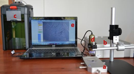
Figure 2: MHT equipment for in-situ analysis of shrinkage activity. (a) micro heating plate (Linkam LTS 120); (b) temperature controller; (c) water circulator; (d) digital microscope; (e) computer with imageMHT software.
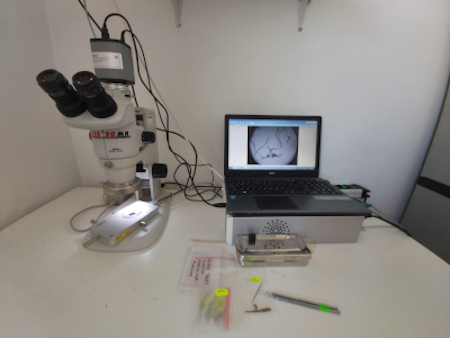
Figure 3: MHT equipment for laboratory analysis of shrinkage activity: micro heating plate Linkam LTS 120 with temperature controller, benchtop stereo microscope with digital camera and computer with imageMHT software.
Another advantage of the MHT method is that it only requires a few tiny fibers (0.1–0.2 mg) which are placed on a microscope slide with a concavity, completely immersed in demineralized and degassed water to ensure thorough wetting. The fiber bundles are well separated under the microscope using fine needles. Then, the microscope slide, covered with a cover glass, is placed on the Peltier element of the heating plate and heated at a rate of 2 C·min−1 in the range (25–95) C. The collagen fiber motion is recorded and the shrinkage intervals evaluated.
Conclusion
Preservation techniques for ancient animal fiber-based artifacts such as leather and parchment are essential in order to maintain the condition of these samples for study, education and display purposes. A better understanding of the aging process and the development of techniques to protect against age-based deterioration are therefore essential. If researchers are able to better understand how collagen behaves in a particular artifact, it is possible to gain helpful insight into its manufacture, how damaged it is, as well as how best to formulate conservation treatments and apply appropriate storage and handling conditions to preserve it.
In order to understand these materials, researchers at ARCH Laboratory have been extensively using the Micro Hot Table (MHT) method since 2006 and are now strongly advocating a more analytical use of this method when analyzing both new and artificially aged samples, and ancient materials, by considering the analysis of all shrinkage parameters and performing complementary non-destructive analysis to limit the risk of over or underestimating the conservation condition of parchment and leather.
References
1. R. Larsen et al., Journal of Society of Leather Technologists and Chemists 1993;77:151.
2. R. Larsen, Termochimica Acta 2000; 365:85.
3. E. Badea et al., e-Preservation Science 2012;9:97
4. C. Carsote, and E. Badea, Heritage Science 2019;7, 48
5. Differential Scanning Calorimetry (DSC), Thermomechanical Analysis (TMA), Dynamic Mechanical Analysis (DMA)